What is Radiomics?
Turning medical images into data

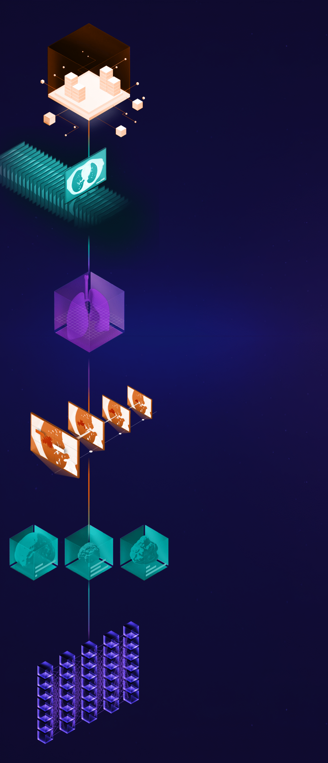
Linked to secure storage archives, the platform streamlines image collection and centralized image quality assessment for enhanced efficiency.
Combining automatic AI image segmentation models and manual reviews into an efficient workflow, accurately identifying and tracking regions of interest (ROI) over time, and integrating both pre- and post-processing tasks, the platform ensures full traceability and auditability of imaging processing steps.
As a single point of control for the management of image feature quantification toolbox, the platform provides optimal and efficient delivery of results for analysis. Ikonics provides a real-time comprehensive overview of each stage of the radiomic pipeline ensuring quality, control, and a full audit trail.

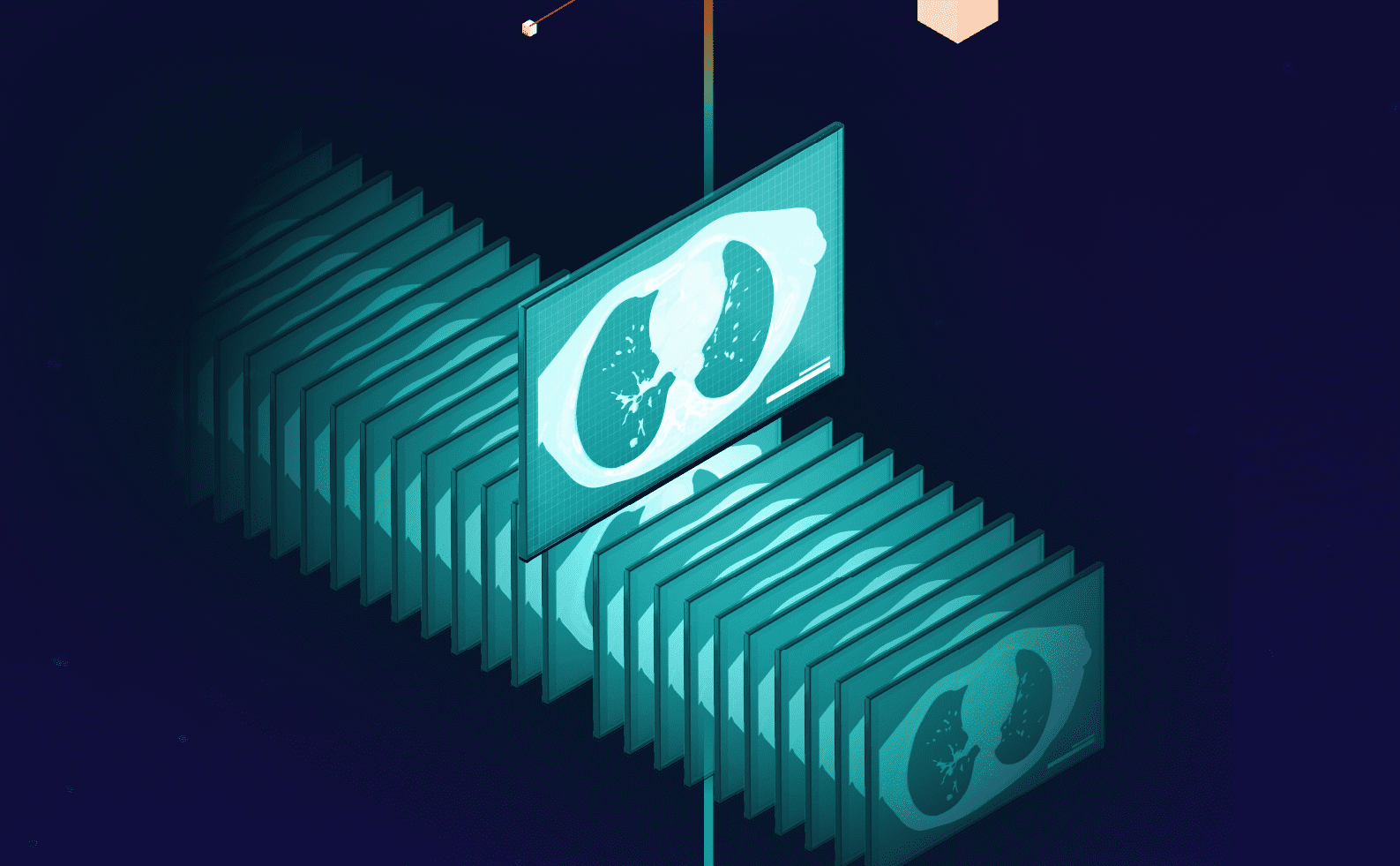

Images are collected and stored on a secure platform based on secure File Transfer Protocol (sFTP), these images are then optimally organized for efficient and accurate processing.
Linked to secure storage archives, Ikonics streamlines image collection and allows us to characterize and structure medical images for every patient according to:


The quality of medical images is assessed based on eligibility criteria conducive to effective analysis.
Building a comprehensive, structured and readable imaging database is required to correctly process data. Linked to a secure storage archive, Ikonics® consolidates the image collection process and centralizes image quality assessment for enhanced efficiency. Perfect characterization and assessment are vital to defining what analysis and insights can be derived from each element of data. During this step we perform:
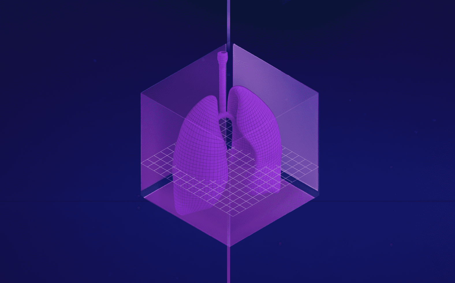
Imaging segmentation is the delineation of the region of interest, portioning the pixels in an image into district regions to assist in image processing tasks. This process can be manual, automated or semi-automated.
The segmentation process is one of the most critical steps of radiomic analysis. To ensure the process is completed to the highest standard, we have an internal team of annotators and medical professionals, as well as a network of radiologists, experts and privileged partners. Furthermore, all segmentations at radiomics.bio are validated by our medical team through an intensive quality assessment process (human-in-the-loop).
During this step, Ikonics® allows us to combine automatic AI image segmentation models and manual reviews into an efficient workflow, accurately identifying and tracking regions of interest (ROI) over time.
These ROIs can include whole organs, oedemas, necrosis, vessels, and abnormalities such as reticulation, ground glass opacity in the lung and diffused disease. Ikonics® also integrates both pre- and post-image processing tasks, giving complete control of the image feature quantification process for an optimal and efficient delivery.
The lesions present in scans are given consistent labels to ensure accuracy. Lesions are linked with different timepoints in a process called “lesion tracking,” in which semantic links are established between regions of interest.
Lesion tracking is the linking of lesion present in different timepoints. With Ikonics® we can accurately identify and track regions of interest (ROI) over time. Consistent labels are given to the lesions present in the scans to ensure accuracy.
The inclusion of relevant radiological findings is also performed at this stage which is incorporated into patient specific reports for a comprehensive assessment of the medical image. Finally, post-processing steps are implemented to ensure perfect quality masks for the quantification of imaging features.
Radiomic features are extracted from the medical scans, each linked to specific characteristics describing the ROI over time in relation to response or lack thereof to treatment, Radiomic features belong to different categories, each of them quantitatively describes single characteristics of organs or lesions under examination, representing the size, shape, texture, and intensity of the ROI. Radiomic signatures are a combination of radiomic features used inside statistical models to predict or classify a specific condition or clinical endpoint of interest.
In this step radiomic features are extracted and quantified in order to describe the ROI at baseline and at multiple timepoints to understand how the lesion evolves following treatment. For research and healthcare applications, radiomics.bio uses machine learning algorithms to ensure explainability of results and provide confidence on the underlying biological mechanisms at play.
The final step in the radiomic workflow converts statistics to insights, ensuring that the quantified statistics are in line with biology.
We provide a wide range of deliverables with a variety of levels of complexity:
We review the relevance of the results obtained and how it correlates to biology including how the results relate to drug mechanisms, patient indications, the localization of the tumor and tumor mutations etc., providing actionable information for insight-based decision-making.

Ikonics® ensures robust, auditable and fully traceable seamless storage and management of advanced AI image analysis.

Centralized, secure image storage with automated quality control for reliable, regulatory-compliant data processing.

Advanced segmentation and radiomic feature biomarker analysis to quantify disease and effect fueling treatment response predictions.
Regular quality control checks by our professionals

Rapid imaging analytics capabilities to optimize clinical trial assessment, improve patient stratification and enhance regulatory submissions.
Ikonics® is a fully integrated proprietary imaging platform managing our image treatment workflow and allows radiomics.bio’s experts to perform each processing step seamlessly and reliably. The Ikonics® platform enables delivery of fast, reliable and impactful results compliant with industry standards.
Segmentation is the identification and delineation of the Regions of Interest(s)(ROI) in medical scans. In this process the Region of Interest (cancer lesions, lesions, organs, lymph nodes etc.) are highlighted to allow for the measurement of the relevant features (such as volume or other radiomic variables) and their evolution can be tracked over time as a result of treatment. This provides an insight into the effect the therapeutic is having on a lesion, a patient or a population as well as changes in the microenvironment. At radiomics.bio, segmentations are typically semi-automatically with all automatically segmented scans verified and if needed, corrected by internal radiologists.
Patient stratification aims to tailor medical treatments to patients based on their own specific characteristics. Radiomics features total tumor burden and volumetric analyses provides comprehensive profiles of patients allowing you to stratify patients based quantitative imaging biomarkers and tumor morphology. This will allow you to differ patients who are likely to respond from those who are resistant and select the best therapeutic option.
Radiomics converts medical images into statistically interpretable and quantifiable data not visible to the human eye. This information can be used to characterize disease, describe and evaluate response, predict clinical outcomes and identify the patients who are most likely to respond to treatment. With radiomics you can optimize your clinical trials by enhancing your understanding of the disease on a patient, organ or lesion level, explore and validate drug mechanisms, obtain early insights and quantify response for objective decision making during each phase of clinical development.
At radiomics.bio we are the pioneers of the science of radiomics. We treat every project as if it was our own and we are dedicated to understanding your needs. At radiomics.bio we focus on using science to provide an answer to a clinical development question. Our objective is to deliver value and allow for insight-based decision making. At radiomics.bio, our only goal is to ensure the success of your clinical study.